Super Resolution
Multi-modal image super resolution and particle size analysis


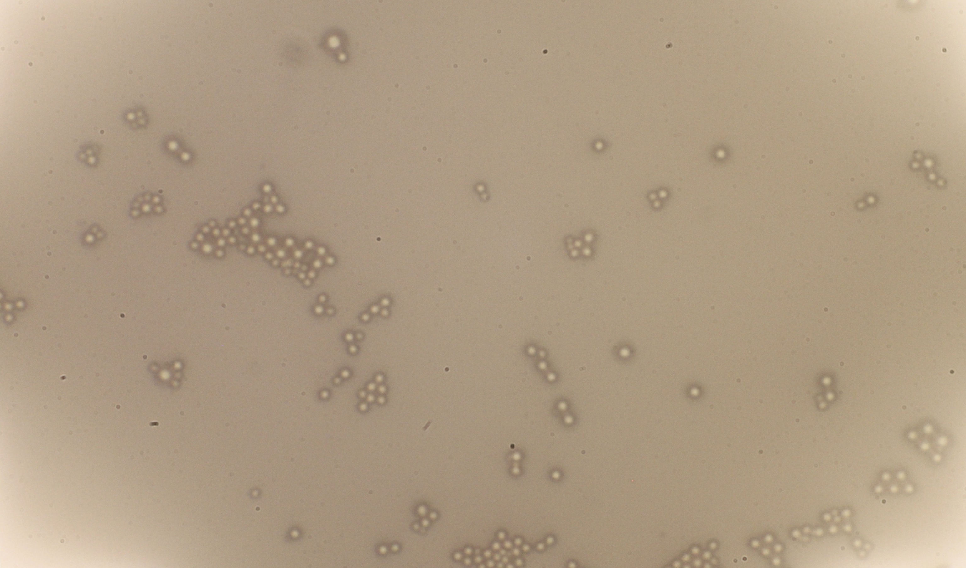
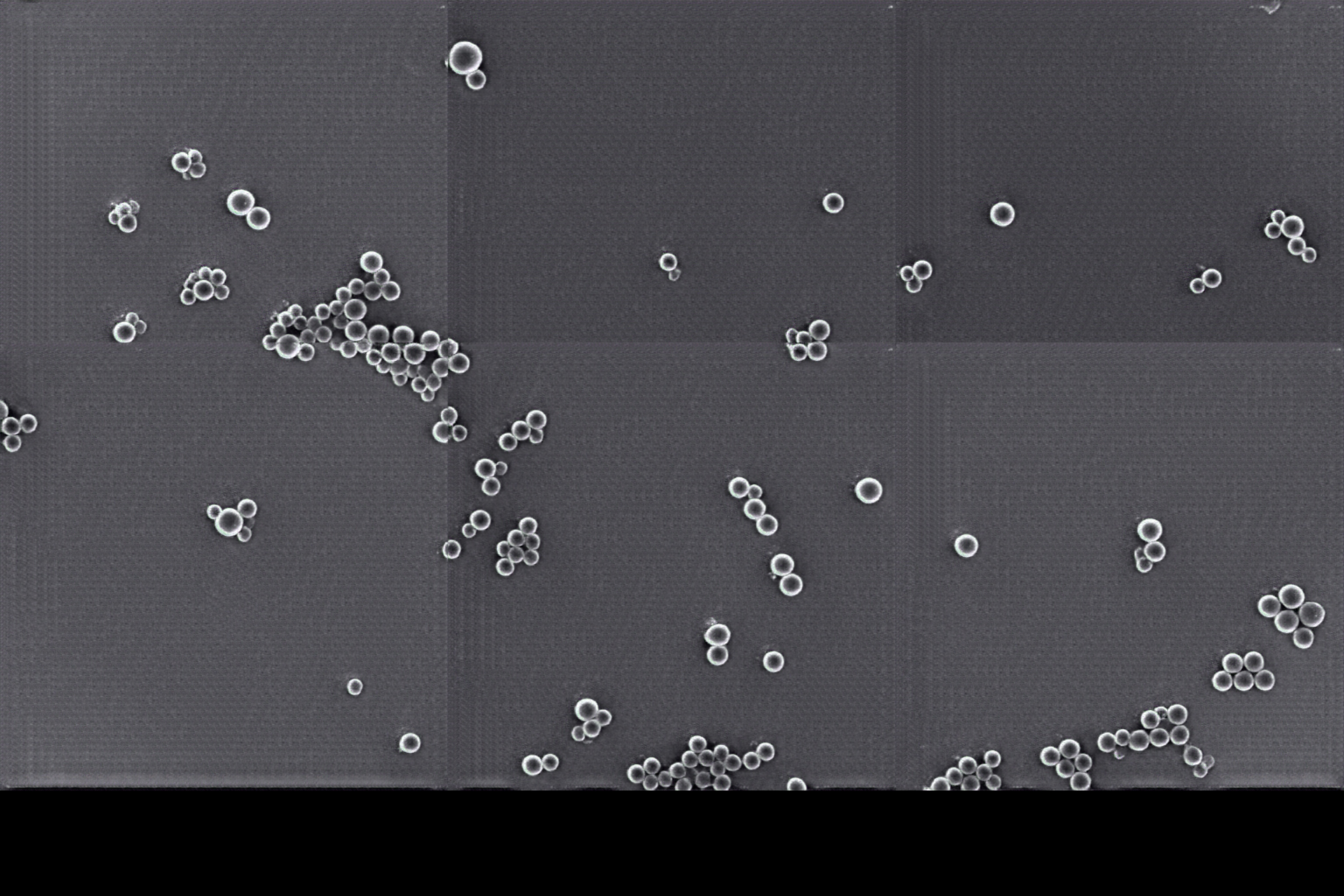
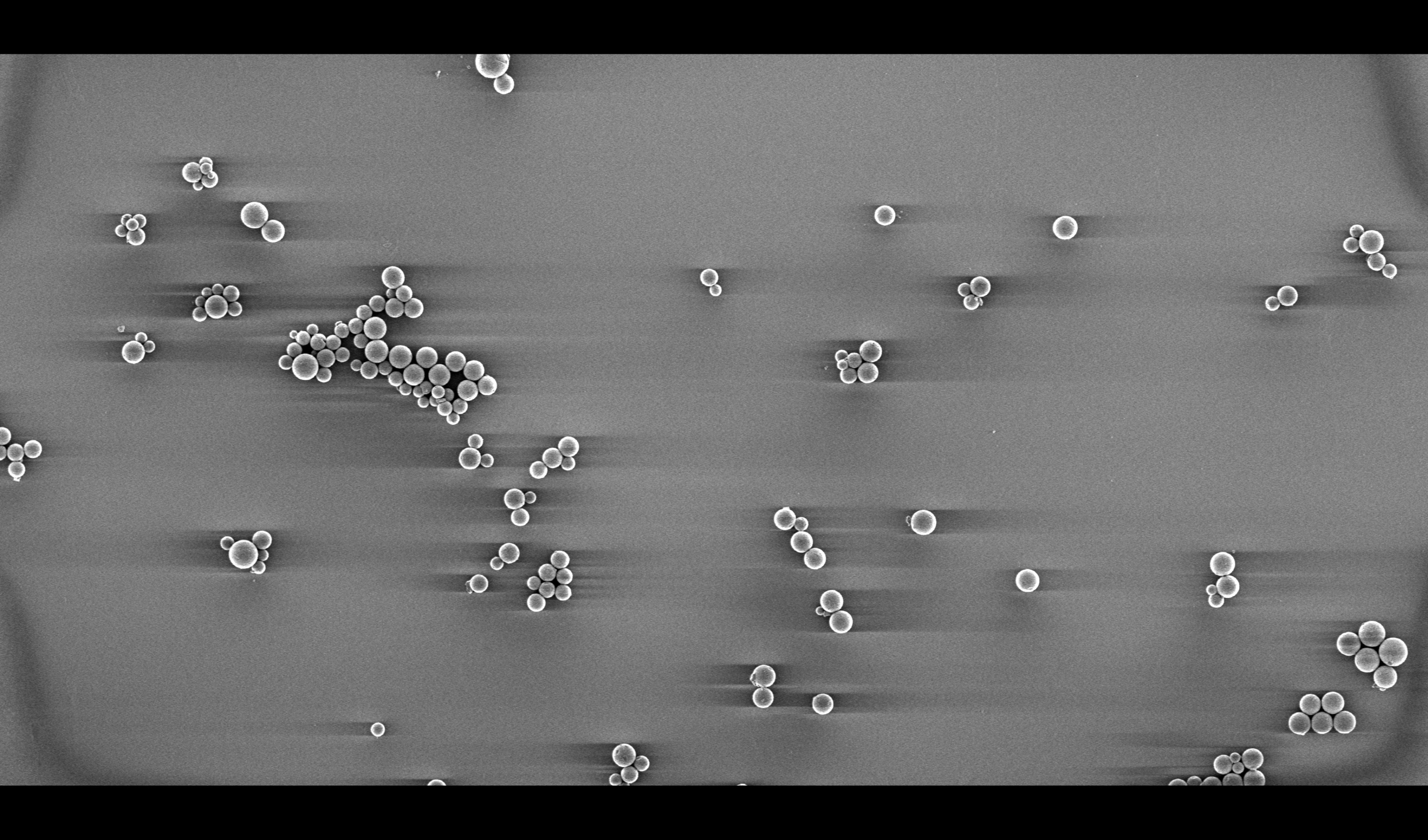
This project was used in an R&D pipeline, which involved accurate sizing of microscopic particles. Optical Microscope images were given as input. Super Resolved images that look like SEM were generated by a combination of AASNA-Pix2Pix and an ESRGAN with Spectral Normalization.
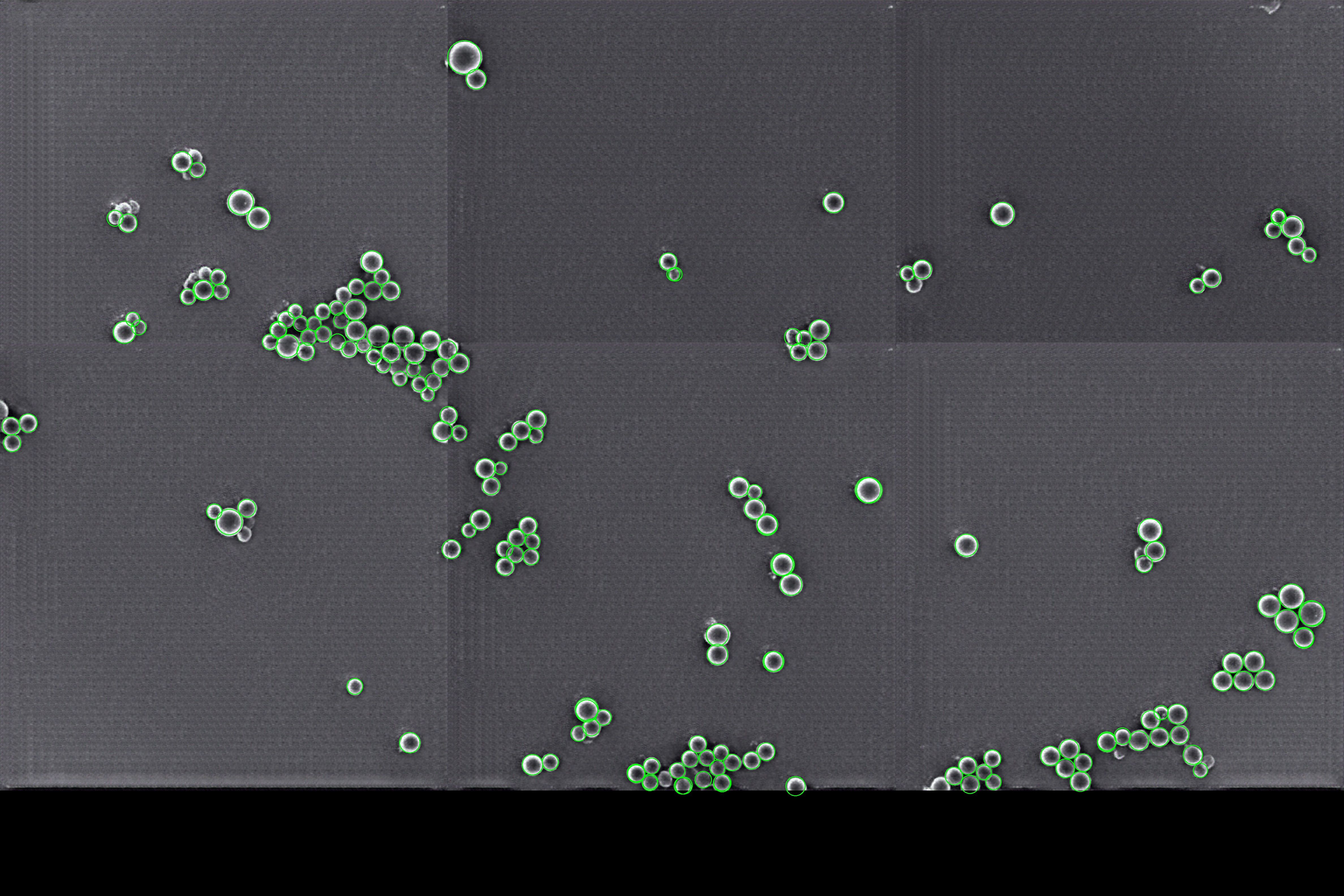
Further, a custom object detection model was created for the super resolved images: Super Resolution Particle Sizing Algorithm. The detection model gives output feature maps as 4D-vectors, each pixel giving a map of (p, x, y, r) to detect the circle of a particle and its radius.

The object detection model for highly agglomerated microscopic particles achieved 0.989 D50 and 0.982 CV values with respect to expert human annotators. The model was deployed in production as a REST API for a battery manufacturing company in Silicon Valley. (name undisclosed due to NDA)